Back Muscles Diagram - Lower Back Muscle Anatomy And Low Back Pain / Muscles of the back diagram with lower back anatomy.. The muscles of the lower back help stabilize, rotate, flex, and extend the spinal column, which is a bony tower of 24 vertebrae that gives the body structure and houses the spinal cord.the spinal. The back has a total of 40 muscles. See back muscles and low back pain. These structures work together to support the body, enable a range of movements, and send messages from the. Daniel nelson on january 1, 2019 2 comments 🔥!
For more information visit www.threetreasuresstudio.com The back has a total of 40 muscles. The following diagram below is the diagrams of back muscle. Muscle anatomy anterior view 12 photos of the muscle anatomy anterior view muscle anatomy anterior view, shoulder muscle anatomy anterior view, human muscles, muscle anatomy anterior view, shoulder muscle anatomy anterior view The back muscles can be three types.
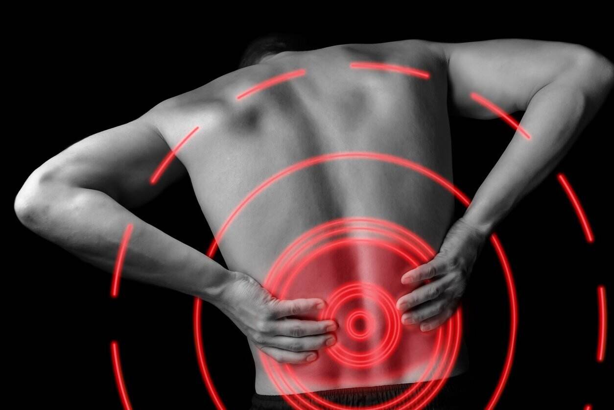
Lower Back Muscle Anatomy And Low Back Pain from marvel-b1-cdn.bc0a.com A strain can be an injury to a tendon attachment from muscle to bone. The latissimus dorsi, also known as the lats or wings, are. Muscle spasms (contraction or stiffening of the back muscles) muscles that feel tight; Human musculature bodybuilding infographic muscular system vector human anatomy back muscle anatomy bicep male muscular anatomy human body anatomy female female anatomy muscle hamstrings muscle. There are several different layers of muscles in your back that are often pulling in different and various directions. The back muscles can be three types. This muscle is a major generator of lower back and hip pain, as well as being responsible for complaints of a burning sensation along the posterior superior iliac spine (psis) and sacroiliac joint. When back development is the goal, stick to one of these variations.
Intermediate back muscles and c.
There are several different layers of muscles in your back that are often pulling in different and various directions. The extrinsic (superficial) back muscles, which lie most superficially on the back. Related posts of back muscle diagram & pain muscle anatomy anterior view. The back has a total of 40 muscles. The latissimus dorsi, also known as the lats or wings, are. We hope this picture anatomy of back muscles diagram can help you study and research. Others, like sumo deadlifts, have been shown in emg studies—and in the trenches—to focus more on other muscle groups than the back. Muscles of the back diagram with lower back anatomy. We think this is the most useful anatomy picture that you need. These muscles include the large paired muscles in the lower back, called erector spinae, which help hold up the spine, and gluteal muscles. This is a diagram of the larger and more surface muscles of the low back. Muscle spasms (contraction or stiffening of the back muscles) muscles that feel tight; Five pairs of lumbar spinal nerves labeled l1 to l5 branch off your spinal cord and exit through small holes between the vertebrae.
Another common cause of lower back and hip pain is disc injury. Symptoms of muscle pain include: Anatomy of the spine and back spine muscles diagram. Human musculature bodybuilding infographic muscular system vector human anatomy back muscle anatomy bicep male muscular anatomy human body anatomy female female anatomy muscle hamstrings muscle. The muscles of the lower back help stabilize, rotate, flex, and extend the spinal column, which is a bony tower of 24 vertebrae that gives the body structure and houses the spinal cord.the spinal.
Low Back Muscles Anatomy Anatomy Drawing Diagram from i.pinimg.com A strain can be an injury to a tendon attachment from muscle to bone. For more information visit www.threetreasuresstudio.com The part of the nerve that emerges out of the spine is called the nerve root. This muscle is a major generator of lower back and hip pain, as well as being responsible for complaints of a burning sensation along the posterior superior iliac spine (psis) and sacroiliac joint. The back consists of the spine, spinal cord, muscles, ligaments, and nerves. The muscles of your back support your spine, attach your pelvis and shoulders to your trunk, and provide mobility and stability to your trunk and spine. In this image, you will find 1st cervical vertebrae, atlus, cervical plexus, 7th cervical vertebrae, 1st thoracic vertebrae, brachial plexus, spinal dura mater, filaments of spinal nerve roots, 12th thoracic vertebra, 1st lumber vertebra, iliohypogastric nerve, ilioinguinal nerve, lumbar. The muscles of the back are a group of strong, paired muscles that lie on the posterior aspect of the trunk they provide movements of the spine, stability to the trunk, as well as the coordination between the movements of the limbs and the back muscles are divided into two large groups:
The deep back muscles, also called intrinsic or true back muscles, consist of four layers of muscles:
The deep back muscles, also called intrinsic or true back muscles, consist of four layers of muscles: Muscle strain is often the cause of back pain from heavy lifting or vigorous exercise. This is a diagram of the larger and more surface muscles of the low back. See back muscle anatomy stock video clips. These muscles include the large paired muscles in the lower back, called erector spinae, which help hold up the spine, and gluteal muscles. Three types of back muscles that help the spine function are extensors, flexors and obliques. The pelvis at the bottom of the back and the shoulders at the top of the back give the back. The back anatomy includes the latissimus dorsi, trapezius, erector spinae, rhomboid, and the teres major. The muscles of the back that work together to support the spine, help keep the body upright and allow twist and bend in many directions. Daniel nelson on january 1, 2019 2 comments 🔥! For more anatomy content please follow us and visit our website: The human back extends from the buttocks to the posterior portion of the neck and shoulders. The muscle elevates, depresses, rotates, and retracts the scapula, or shoulder blade.
The muscles of the lower back help stabilize, rotate, flex, and extend the spinal column, which is a bony tower of 24 vertebrae that gives the body structure and houses the spinal cord.the spinal. It is opposite from the chest, and the vertebral column runs down the back. Muscle anatomy anterior view 12 photos of the muscle anatomy anterior view muscle anatomy anterior view, shoulder muscle anatomy anterior view, human muscles, muscle anatomy anterior view, shoulder muscle anatomy anterior view Daniel nelson on january 1, 2019 2 comments 🔥! We hope this picture anatomy of back muscles diagram can help you study and research.
Back Workout To Build A Lean Sculpted V Shaped Back A Lean Life from aleanlife.com Back muscles, back muscle diagram. A strain can be an injury to a tendon attachment from muscle to bone. Nerves in your lower back. Muscle spasms (contraction or stiffening of the back muscles) muscles that feel tight; Muscle anatomy anterior view 12 photos of the muscle anatomy anterior view muscle anatomy anterior view, shoulder muscle anatomy anterior view, human muscles, muscle anatomy anterior view, shoulder muscle anatomy anterior view Three types of back muscles that help the spine function are extensors, flexors and obliques. Lower back muscle diagram anatomy does degenerative disc disease affect the lower back muscle? The muscles of your back support your spine, attach your pelvis and shoulders to your trunk, and provide mobility and stability to your trunk and spine.
A clip from 3d back muscles:
The muscle elevates, depresses, rotates, and retracts the scapula, or shoulder blade. Anatomy of the spine and back spine muscles diagram. These muscles include the large paired muscles in the lower back, called erector spinae, which help hold up the spine, and gluteal muscles. This muscle is a major generator of lower back and hip pain, as well as being responsible for complaints of a burning sensation along the posterior superior iliac spine (psis) and sacroiliac joint. Three types of back muscles that help the spine function are extensors, flexors and obliques. We think this is the most useful anatomy picture that you need. Support and protect your spine; The extrinsic (superficial) back muscles, which lie most superficially on the back. The muscles of the abdomen, lower back, and pelvis are separated from those of the chest by the muscular wall of the diaphragm, the critical breathing muscle. The muscles of your back support your spine, attach your pelvis and shoulders to your trunk, and provide mobility and stability to your trunk and spine. Superficial back muscles, intermediate back muscles and intrinsic back muscles.the intrinsic muscles are named as such because their embryological development begins in the back, oppose to the superficial and intermediate back muscles which develop elsewhere and are therefore classed as extrinsic muscles. Creatine research more than a sports supplement read more…. Superficial, intermediate, deep and deepest layers.these muscles lie on each side of the vertebral column, deep to the thoracolumbar fascia they span the entire length of the vertebral column, extending from the cranium to the pelvis